Dermoscopy: What Is It And What Is It For?
The use of dermoscopy has offered a new dimension to skin lesions and has provided an effective diagnostic tool for differentiating benign or malignant skin tumors. In the same way, it also supports the correct identification in many other pathologies of general dermatology.
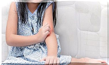
The skin diagnosis is often, but not always, based on vision. Dermatologists tend to find situations in which the options may be several, which makes it difficult to determine if you are dealing with a lesion of one type or belonging to another classification.
What is dermoscopy?
Dermoscopy, also known as epiluminescence microscopy or skin surface microscopy , is a non-invasive technique performed on living patients. In other words, it is a practice that takes place in the offices designed and specialized for this purpose.
Traditionally, it has found frequent use in the evaluation and differentiation of melanocytic lesions suspected of melanomas, as well as skin cancers. Among the latter we can mention basal cell carcinoma and squamous cell carcinoma.
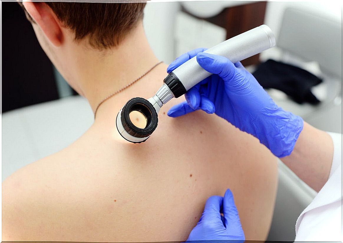
The dermatoscope as an instrument
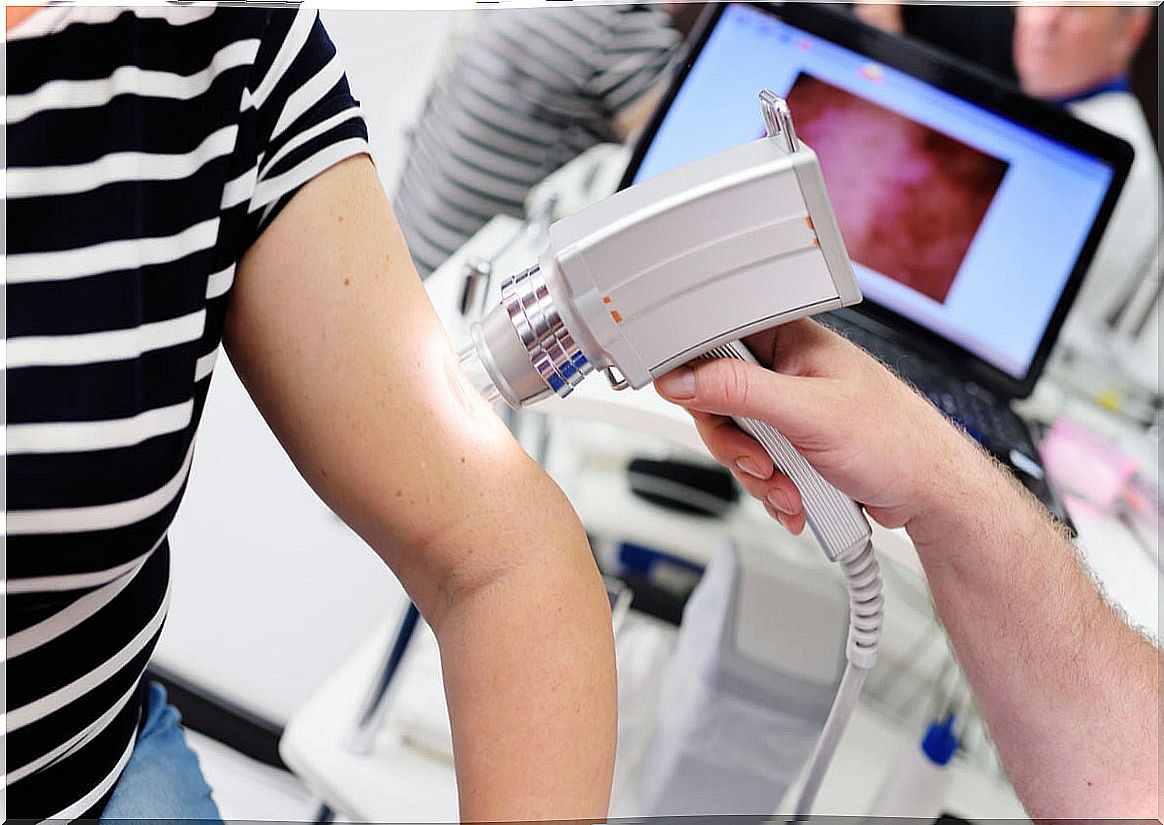
A dermatoscope is the device used to carry out this diagnostic method. It can assess structures down to the depth of the reticular dermis and record images for future comparison, just like photographs.
The essential components of this type of device are the following:
- Achromatic lenses with magnification ranging from 10X to 200X or even more.
- A built-in lighting system comprised of halogen lamps placed inside the handpiece.
- A power source: these can be rechargeable or replaceable batteries.
You may be interested: Hyperpigmentation: why does it happen?
What is dermoscopy for?
The basic principle of dermoscopy is the transillumination of a lesion to study it under high magnification. In this way, subtle features can be visualized that would go unnoticed. The technique makes it possible to reveal colors and microstructures not visible to the naked eye that correspond to attributes of the tissues.
Findings should always be interpreted within the general clinical context of the patient. The image is integrated with the information from the clinical history and the macroscopic examination.
Applications for this detailed examination of the skin surface are being discovered in a few other circumstances, such as the following:
- Unpigmented skin lesions.
- Infectious dermatitis.
- Nail disorders.
- Scabies mites inside a tunnel in the skin.
- Locate a splinter.
- Exam of the capillaries of nail fold in lupus erythematosus cutaneous (LES) or the sclerosis systemic (ES).
- Distinguish certain skin conditions, such as lichen planus, from others such as psoriasis or eczema.
- The evaluation of hair loss, under the name of trichoscopy.
According to studies published in Dermatology Practical and Conceptual , scabies is, without a doubt, the skin disease whose diagnosis has benefited the most from the use of the dermatoscope. This without considering oncological pathologies.
Types of dermoscopy
According to the light source used by the dermatoscope itself, today two types of dermoscopy can be distinguished, which are the following:
- With conventional non-polarized light (NPD) : in these devices, a drop of oil must be applied to the lesion to be analyzed to avoid the phenomenon of light scattering that makes visual examination difficult.
- Contact with polarized light (PCD): these devices have a special, polarized light that cancels the scattering phenomenon and, therefore, does not require immersion oil on the lesion. The device can directly contact the lens with the skin surface.
Some dermatoscopes have a built-in photography system, with support software for image capture and storage. For those who do not have this mechanism, there are special adapters available to connect to digital cameras.
Read also: Benign skin tumors: how do they manifest themselves?
How is it done and what is its preparation?
Dermoscopy is a simple procedure that is performed at the time of consultation with the dermatologist. The patient must expose the lesion to be analyzed.
The dermatoscope will be supported on the area to visualize the corresponding morphological characteristics. It is a painless procedure that does not generate discomfort or complications.
No particular preparation is required, but it is recommended to go to the doctor’s office with comfortable clothing and with the surface of the skin to be evaluated clean and without creams. Once the dermoscopy is complete, the person can return to their usual activities without any further indication.
Dermoscopy as a complement
Dermoscopy has far-reaching uses beyond the simple diagnosis of skin, hair, and nail disorders. New studies reveal that its applications exceed the oncological and that it is possible that it serves as a support for infections and parasitosis.
The findings are highly relevant, both for physicians and patients. They offer a quick resolution to diagnostic and therapeutic dilemmas that are routinely encountered.